Türkçe
 Русский
 Deutsch
Laser Capillary Treatment
Laser treatment for capillaries is an effective method for eliminating fine vascular dilations (telangiectasias) on the face and legs. Laser energy targets hemoglobin inside the vessel, generates heat, and controllably injures the vessel wall. As a result, the vessel closes and the skin’s appearance improves.
For capillary problems, laser therapy is particularly suitable for superficial and small-caliber vessels. Wavelength and energy level are adjusted according to skin type and vessel depth. Surrounding tissues are protected during the procedure, reducing the risk of side effects. The treatment is usually completed quickly without the need for anesthesia.
Advantages of laser vein therapy include minimal or no pain during the session, a high likelihood of scar-free healing, and a rapid return to social life. The number of sessions varies with vessel density; noticeable improvement typically occurs after several treatments. Mild redness or swelling after the session subsides quickly.
Post-treatment care includes sun protection, moisturizing the skin, and avoiding high heat. Proper device selection and expert supervision deliver lasting results and high aesthetic success. Regular follow-ups are important to help prevent the formation of new vessels.
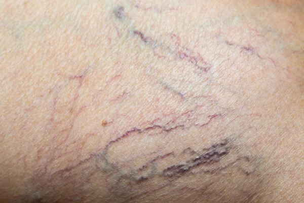
Capillary (spider vein) issues are more common than you might think. A large portion of adults—especially on the legs and face—experience them to varying degrees. Although the exact mechanism behind their appearance is not fully clarified, two main factors are most often considered:
Increased intravascular pressure (venous hypertension). The valves in the leg veins, which return blood to the heart, wear out over time or stop working optimally. Blood then flows backward (reflux), increasing venous pressure. This elevated pressure dilates small vessels near the skin’s surface, making them visible.
Structural weakness of the vessel wall. In some people, genetic predisposition leads to a congenitally weaker vein wall. Hormonal changes (pregnancy, menopause, hormone therapy) and excessive sun exposure can further reduce elasticity and strength.
Common risk factors:
This is a critical point in our specialty. Leg spider veins are not merely a cosmetic concern; they can be the earliest and most “innocent-looking” sign of underlying chronic venous insufficiency (CVI) that you cannot see on the surface.
We must identify the source before treatment. Otherwise, you remove only the visible vessels, but because the underlying high-pressure source persists, the spider veins return quickly (recurrence)—the most common cause of disappointment.
Therefore, venous duplex ultrasound (VDU) is absolutely mandatory before any lower-limb vein treatment. With VDU we assess:
Our priority is not just to erase the visible spider vein but to correct the hemodynamic cause that produced it.
There’s no “magic” in laser therapy—it’s physics, specifically the theory of selective photothermolysis.
In simple terms: the chosen laser energy targets a specific chromophore inside the vessel, heats and closes the vessel while sparing nearby tissue.
Three key components of laser treatment:
Laser light is absorbed by hemoglobin in red blood cells. The absorbed energy turns into heat, coagulating the vessel wall, narrowing it, and leading to durable closure.
The target chromophore appears in two forms:
Oxyhemoglobin — in oxygenated, red blood (e.g., facial vessels).
Deoxyhemoglobin — in darker/more bluish venous blood (e.g., leg veins).
Wavelength determines how deeply light penetrates the skin and which hemoglobin form absorbs it best.
Short wavelengths (e.g., 532 nm): very superficial penetration; strongly absorbed by oxyhemoglobin. Ideal for thin, red facial vessels.
Long wavelengths (e.g., Nd:YAG 1064 nm): penetrate several millimeters; effectively absorbed by deoxyhemoglobin. Essential for deeper, bluish leg vessels.
This is the time the laser energy is delivered. It must be long enough to close the vessel before heat diffuses into surrounding tissue. This depends on thermal relaxation time (TRT). The larger the vessel diameter, the longer the pulse needed—hence millisecond-range long pulses for capillary treatment.

The device is individualized based on location, color, depth of the vessel, and patient skin phototype.
For facial capillaries (red and superficial):
KTP laser (potassium titanyl phosphate, 532 nm):
Strongly absorbed by oxyhemoglobin.
Very effective for superficial lesions.
Commonly used in lighter skin (Fitzpatrick I–III).
Higher burn risk in darker skin due to melanin absorption.
Pulsed Dye Laser (PDL, 585–595 nm):
The “gold standard” for port-wine stains (congenital vascular malformations).
Also used for facial telangiectasias and rosacea.
For leg capillaries (blue and deeper):
Long-pulsed Nd:YAG (1064 nm):
Provides deep penetration.
Indispensable for thicker, deeper reticular veins.
Minimally absorbed by melanin, thus the safest and most effective choice in darker skin (Fitzpatrick IV–VI).
Other versatile systems:
IPL (intense pulsed light):
Not a single laser line but a broad spectrum.
Can address vascular (redness) and pigmented lesions simultaneously.
Effective on the face, but use cautiously in darker skin due to post-inflammatory pigmentation risk.
A very common question. The simple answer: both! The best results often come from a combined approach. Selection is diameter-based—an algorithm is used:
Treatment by diameter:
Sclerotherapy (foam or liquid) is superior and more efficient. As diameter increases, a sclerosant closes the vein better than laser.
When laser has an edge:
Optimal combined strategy:
First correct any truncal (axial) venous insufficiency, if present. Then close the larger blue “feeder” reticular veins with sclerotherapy. Finally, clear the remaining finest red networks with transdermal laser. This yields the highest efficacy and durability.
Safety and success depend on preparation. Key rules before therapy:
Outcomes depend greatly on device quality and operator experience. Two fundamental principles:
Cooling prevents surface burns and reduces discomfort. Methods include:
Dynamic Cooling Device (DCD): a cryogen spray just before the pulse.
Contact cooling: a chilled sapphire window on the handpiece.
Application of cooling gel.
The clinician selects fluence and pulse duration (ms) according to vessel diameter, depth, and skin phototype. Critical points:
Avoid pulse stacking (overlapping): this causes excessive heat buildup and burns. Leave ~1 mm spacing between pulses.
Therapeutic endpoint: visually, immediate blanching, disappearance, or “collapse” of the vessel (photocoagulation). Whereas purpura was once the target, modern lasers aim for vessel closure without bruising.
The first few weeks shape the final result.
Immediately after: vessels may look dark red/purplish with swelling and mild erythema. Note: vessels don’t vanish “instantly”; fading takes 2–6 weeks, sometimes longer.
Pain: usually mild; managed with OTC analgesics (often acetaminophen).
Mandatory recommendations:
Walk 30–60 minutes immediately after.
Continue at least 30 minutes daily thereafter.
Critical to prevent thrombosis and stimulate circulation.
Avoid hot baths, saunas, steam rooms, and pools for 1–3 weeks.
Heat induces vasodilation and impairs healing.
Avoid heavy exercise (running, weightlifting) for 1–2 weeks.
Shield the treated area from sun for at least 4 weeks and use high-SPF sunscreen.
Laser therapy has a high safety profile, but side effects/complications can occur. Most are transient and often relate to incorrect parameters.
Common, reversible effects:
Significant complications:
Hyperpigmentation: the most common after laser/sclerotherapy; due to hemosiderin deposition from residual blood in the closed vessel. Usually resolves spontaneously or with pigment-targeted care.
Hypopigmentation: less common—areas of lightening.
Excess epidermal heating may cause blisters, crusting, and persistent scars. Nearly always due to improper settings or pulse overlap.
Very fine new red vessels around the treated area. Often self-limited; occasionally needs additional treatment.
Small firm clots in treated superficial veins.
If your capillary treatment is performed by a specialist who also corrects truncal venous insufficiency (e.g., EVLA), long-term outcomes are excellent.
Truncal vein closure (EVLA): success at 12 months is ~98% in axial veins—indicating durable correction of the root cause.
Recurrence risk: reopening of the treated vessel is uncommon. However, venous disease is genetically influenced and progressive, so new areas of insufficiency may appear over time. Even at 5-year follow-up, recurrence rates remain acceptable.
Remember: capillary treatment is not a one-off “fix” but the management of a chronic condition requiring periodic checkups and lifestyle adjustments. Regular follow-ups are key to long-term success.
If leg spider veins bother you cosmetically and you also experience any of the following, consult a vein specialist:
These signs suggest possible chronic venous insufficiency—laser alone will not be sufficient in that case.
Laser can be applied to the face, around the nose, legs, and chest—areas where fine vessels are common. It’s effective for reducing the appearance of red-purple vessels that cause cosmetic concern.
Laser light is absorbed by hemoglobin and converted to heat, which controllably injures the vessel wall. The vessel is then gradually resorbed by the body.
Mild tingling or burning may occur, but most patients tolerate it well. Cooling systems and anesthetic creams enhance comfort.
Yes. Temporary redness or minor bruising is common and usually resolves within a few days. The skin soon returns to normal.
It depends on the density and thickness of vessels. Most patients see noticeable improvement within 1–5 sessions spaced 4–6 weeks apart.
The same vessels do not “return,” but new ones may develop in similar areas. Sun protection, quitting smoking, and regular skin care help reduce new formation.
Avoid hot showers, sauna, and intense exercise for 24 hours. Use sunscreen and keep the skin moisturized to support healing.
It’s not recommended for those with open wounds, recently tanned skin, or those taking photosensitizing medications. It’s generally postponed during pregnancy.
Once treated vessels are fully closed, results are considered long-lasting. However, hormonal factors or genetics may lead to new vessels over time.
Light makeup is acceptable after 24 hours. If redness or irritation persists, wait longer and allow the skin to recover.

Varicose vein treatment prices are determined directly according to a treatment plan tailored specifically for [...]
Read More ➜
A successful varicose vein treatment depends on two critical phases. The most important step before [...]
Read More ➜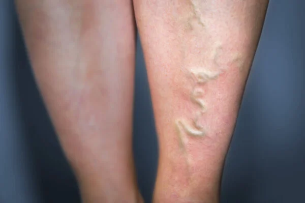
After varicose vein treatment, there is a risk of recurrence, but this risk can be [...]
Read More ➜