
 Türkçe
 Русский
 Deutsch
Sclerotherapy
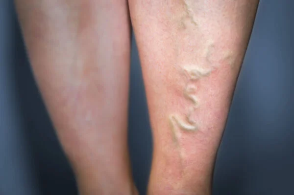
Sclerotherapy and foam treatment are injection-based, minimally invasive methods that close varicose veins. A sclerosing agent in liquid or foam form is injected into the vein; by creating controlled inflammation in the vein wall, it leads to luminal closure. As a result, the appearance of varicose veins diminishes and circulation is regulated.
Foam sclerotherapy is particularly effective in treating medium- and large-caliber superficial veins. The foam form increases the contact time with the inner surface of the vein, enhancing efficacy. When performed under ultrasound guidance, the accuracy of injection increases and the risk of complications decreases. The procedure generally does not require local anesthesia.
Advantages of sclerotherapy include rapid recovery, low cost, and successful cosmetic outcomes. Patients can usually return to daily activities in a short time after treatment. Regular use of compression stockings supports vein closure and increases the durability of the treatment.
Mild redness or bruising may be seen in the post-treatment period; however, these effects are temporary. Regular follow-up and ultrasound monitoring are important for preventing the development of new varicose veins. When performed by an experienced physician, sclerotherapy and foam treatment offer a safe and effective option for venous insufficiency.

The aim of sclerotherapy is to permanently close the problematic vein from the inside by irritating it, rather than surgically removing it. This is done with a special medication called a “sclerosant.” The medication is delivered directly into the problematic vein using a very fine needle.
The drug comes into contact with the innermost layer of the vein—the thin membrane we call the endothelium. This contact creates an intentional, controlled injury in the vein wall. The body immediately initiates a healing process. This process causes the vein walls to stick together and fuse permanently. At the end of the treatment, the problematic vein turns into a fibrous cord that can no longer carry blood. Over time, the body reabsorbs and eliminates this nonfunctional remnant.
The medications that achieve this closure are generally called “sclerosants.” They are classified into different groups according to their mechanism of action. In modern practice, the agents most commonly used—and recommended by European guidelines—are those in the “detergent” group. The main types are:
Foam treatment in particular is based on detergent-group agents (POL and STS).
Foam treatment is one of the most important advances in sclerotherapy. It is the transformation of the liquid sclerosant into “micro-foam” by mixing it with air at a certain ratio (typically 1 part drug to 4 parts air). This is prepared within seconds using a simple technique called the “Tessari method,” with two syringes and a three-way stopcock. Or, today, it can be prepared with devices such as Varixio.
So why go to the trouble of turning a liquid into foam? The answer lies in the physical properties of the foam.
When you inject a liquid drug into a vein, it immediately mixes with blood and becomes diluted. Its concentration falls and its effect decreases.
Foam, however, is much denser and does not mix with blood. When injected, it pushes blood ahead of it like a piston and clears the vein of blood. In this way, the drug remains at full concentration, without dilution, and stays in contact with the vein wall for much longer. This multiplies the effectiveness of the treatment.
Yes—especially in certain vein types, the difference is very clear. Scientific studies and meta-analyses have demonstrated this difference unequivocally.

Sclerotherapy is a versatile treatment that can be used for all venous problems—from the simplest telangiectasias (C1) to the most advanced form, venous leg ulcers (C6). The main conditions that can be treated include:
This is one of the most critical decisions for treatment success and depends entirely on the diameter and type of the target vein. International guidelines draw this distinction very clearly.
In short, apart from cosmetic spider veins, if there is true venous insufficiency and varicose disease in the leg, foam treatment is the preferred method.
This is the “gold standard” and a nonnegotiable rule of modern varicose vein treatment. Color Duplex Ultrasound (DUS) is not just a simple diagnostic tool to check “is there reflux or not?”—it is the roadmap for the entire treatment plan.
Think of the leg vein system as a tree. Sometimes the bulging varix the patient sees on the leg is only a “branch.” The real problem is a leak in the main “root” vein hidden deep inside (usually the great saphenous vein near the groin), which is not visible to the eye.
What happens if the specialist treats only the visible “branch” without finding this “root” with ultrasound? Because the pressure and reflux at the root continue, the body soon finds a new “branch,” and the varicosity recurs elsewhere.
One of the most important causes of treatment failure and recurrence is precisely this: inadequate ultrasound mapping that fails to correctly identify the true source of reflux. In medicine, this is called a “tactical error.”
Ultrasound shows us which vein is problematic, its caliber, depth, where it begins and where it goes. We determine our treatment strategy (liquid, foam, or laser?) entirely according to this map.
UGFS (Ultrasound-Guided Foam Sclerotherapy, i.e., echosclerotherapy) is the standard method for treating deep, subcutaneous trunks and tributaries not visible on the surface (saphenous veins, branches, perforators). The procedural steps are quite simple for the patient.
This method ensures that the drug is delivered to the correct site and does not escape unnecessarily into other veins. For safety, a certain maximum foam volume (e.g., 10 mL) is generally not exceeded in a single session.
Yes—when applied to the right patient, UGFS is a safe, effective, and durable method. Scientific studies show very high success rates in eliminating axial reflux even at 5-year follow-up.
However, the single most important factor influencing the success and durability of foam treatment is the diameter of the treated axial vein (saphenous vein).
This is exactly why pre-procedure ultrasound is so important. Ultrasound clearly answers the question, “Is this vein suitable for foam, or would thermal methods like laser (EVLA) or radiofrequency (RFA) provide a more durable result?” If the vein diameter is within ideal limits, foam can be at least as effective as laser or radiofrequency—while being simpler and more cost-effective as a first-line option.
This point makes the biggest difference for patient quality of life. Although traditional surgery (stripping the vein) is a durable method in the long term, it requires general or spinal anesthesia, incisions, sutures, and a longer recovery period.
Foam sclerotherapy (and other modern methods such as laser/RFA) offers patient-centered advantages. In one scientific study, the average time to return to normal life and work was found to be 13 days for surgery, but only 1–2 days for sclerotherapy.
Even if a small “touch-up” may be needed 5–10 years later, most patients understandably prefer an office-based treatment that does not require anesthesia and allows them to return to normal life in 2 days—rather than a surgical operation that typically requires 13 days off. This is the essence of the “shared decision-making” process.
Foam is much more potent than liquid. This stronger effect triggers a more pronounced healing (inflammatory) response in the vein wall. That can cause slightly more tenderness, pain, or bruising than liquid therapy in the first few days. However, this is a natural and expected process that indicates the treatment is working, and it is well tolerated by most patients.
Interestingly, despite this initial transient discomfort, patient satisfaction surveys show that foam therapy scores significantly higher than liquid therapy. The reason is simple: patients prefer a clearer, more durable result with fewer sessions—over a short period of initial discomfort.
Sclerotherapy is generally a safe method. Serious complications are extremely rare. The most common side effects are usually temporary, harmless, and localized to the treatment area. These include:
Among these, “hyperpigmentation”—that is, discoloration—is the most common cosmetic complaint. It is an iron (hemosiderin) deposition caused by the breakdown of blood trapped in the closed vein. It is not a burn. It usually clears as the body gradually removes it within 6–12 months, though rarely it may persist.
Superficial thrombophlebitis (vein firmness) is, in fact, part of the treatment—a reaction caused by clot formation within the closed vein; the body resorbs it over time. Serious risks such as tissue necrosis (skin ulcer) or deep vein thrombosis (DVT) are very, very rare.
This is a rare but recognized issue unique to foam therapy. A very small percentage of patients may experience certain transient symptoms within the first few minutes after the procedure (usually 5–15 minutes). These symptoms include:
The cause is “paradoxical embolism.” About a quarter of the general population has a small, congenital opening between the right and left atria of the heart called a Patent Foramen Ovale (PFO), which is usually harmless.
When foam is injected, the micro air bubbles inside travel through the venous system to the right side of the heart and would normally go to the lungs to be safely absorbed and exhaled. However, in people with a PFO, some of these bubbles can pass through this opening to the left side of the heart (the arterial side). From there, they may reach arteries supplying the brain and cause the transient symptoms listed above.
These symptoms are almost always completely transient and do not leave any permanent damage. Because of this risk, it is essential to ask patients about a history of “migraine with aura” prior to the procedure.
There are situations in which the treatment is not appropriate. These are divided into absolute and relative categories.
Absolute contraindications:
Relative situations requiring risk–benefit analysis:
Post-procedure care is at least as important as the procedure itself to guarantee success. The process is quite simple and revolves around two basic rules:
Compression (Compression Stockings): Immediately after the procedure, a compressive bandage or compression stocking is applied to the leg. The purpose is to exert external pressure on the vein whose walls have adhered, to keep it closed, prevent it from filling with blood again, and reduce the risk of recanalization. It also reduces post-procedural pain and edema. Typically, patients are advised to wear it for 7 days during daytime, depending on the protocol.
Ambulation (Walking): Patients are encouraged to get up and walk right after the procedure. Walking activates the calf muscle pump, accelerates blood flow, and prevents clot risk. Prolonged bed rest or standing still is discouraged.
Sclerotherapy and foam treatment are generally used for spider veins, reticular veins, and small superficial varicose veins. The treatment closes the vein wall and redirects blood flow to healthy veins.
Mild bruising, redness, or swelling may persist for a few days after treatment. This is a normal healing process associated with vein closure and usually resolves on its own.
Because hormonal changes during pregnancy affect the venous system, sclerotherapy is generally postponed until after delivery. During breastfeeding, it may be performed with physician approval.
Avoid hot baths, sauna, and strenuous exercise for the first 24 hours after treatment. Using compression stockings speeds up recovery and helps prevent new vein dilatations.
Treated veins close permanently, but new varicose veins can develop. Therefore, a healthy lifestyle, exercise, and weight control are important for long-term success.
Patients can usually return to normal activities the same day. However, it is recommended to avoid prolonged standing and strenuous exercise for a few days.
It is not recommended for individuals with a history of deep vein thrombosis, those with active infections, or those who are pregnant. Eligibility must always be determined by a physician’s evaluation.
Mild pain, burning, or a feeling of tightness is normal. Relief can be achieved with cold compresses and physician-recommended creams. In rare cases of severe pain, consult your doctor.
Spider veins generally disappear within a few weeks. For larger veins, full results may take 1–3 months. Skin appearance improves gradually after treatment.
Regular exercise, avoiding prolonged standing, and using compression stockings reduce the risk of new varicose veins. Weight control is also important.

Varicose vein treatment prices are determined directly according to a treatment plan tailored specifically for [...]
Read More ➜
A successful varicose vein treatment depends on two critical phases. The most important step before [...]
Read More ➜ After varicose vein treatment, there is a risk of recurrence, but this risk can be [...]
Read More ➜