
 Türkçe
 Русский
 Deutsch
Echosclerotherapy
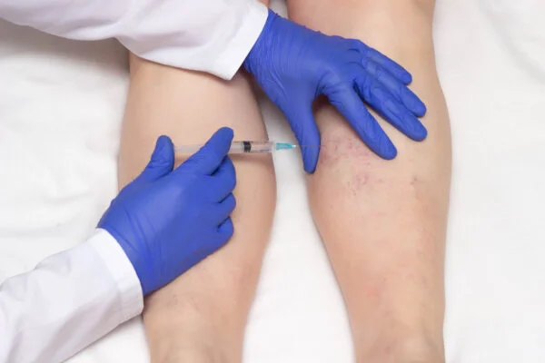
Echo-sclerotherapy is a varicose vein treatment performed under ultrasound guidance. In this technique, a potent sclerosing agent is injected directly into the target vein using ultrasound imaging. The aim is to create a controlled inflammatory reaction in the vein wall to close the lumen of the vessel. In this way, the appearance and symptoms of varicose veins are significantly reduced.
In patients in whom reflux is detected in the deep or superficial venous system, echo-sclerotherapy offers a minimally invasive alternative to surgery. Thanks to ultrasound guidance during the procedure, injection accuracy increases and the risk of complications is minimized. The treatment is usually performed on an outpatient basis and does not require anesthesia.
The advantages of echo-sclerotherapy include rapid recovery, low cost, and cosmetically satisfying results. When the sclerosing agent is used in foam form, its contact time with the vein surface increases, which enhances efficacy. The use of compression stockings after the procedure supports treatment success.
In the post-treatment period, short-term redness or tenderness may be observed; however, these side effects are generally temporary. With regular ultrasound follow-ups, vein closure and the development of new reflux are monitored. When performed by an experienced physician with proper patient selection, echo-sclerotherapy offers a high success rate.
The primary goal of this treatment is to close from the inside a dilated, dysfunctional varicose vein that exhibits backward flow (reflux) instead of surgically removing it. A special medication called a “sclerosant” is used to do this.
This drug acts like a detergent; by disrupting the protein structure of the endothelial cell layer lining the inner surface of the vein, it creates a controlled chemical injury. The body perceives this as a “wound” and immediately initiates a repair process. This process first causes the vein to contract, then its walls to adhere to each other, and finally to transform into permanent connective tissue called “fibrosis.” Blood flow is then automatically redirected to nearby healthy veins.
In the past, sclerotherapy was performed only with the liquid form of the drug. However, this was not very successful, especially in large, high-pressure main veins (such as the saphenous veins). The reason is that the liquid rapidly mixed with blood inside the vein, becoming diluted (reducing its concentration) and quickly neutralized by the blood.
The “foam” form has been a real breakthrough in this treatment. By mixing the drug with air or special gases using a specific technique (the Tessari method), a dense micro-foam is obtained. This foam has tremendous advantages over liquid:
The “Echo” (ultrasound) in the name of the treatment is its most critical component. You can think of ultrasound as our “eyes” in this procedure. Without ultrasound, performing foam therapy in the deeper main veins would be equivalent to a blind injection and could be extremely risky.
Ultrasound has four roles in this treatment.

In modern varicose vein treatment there is no longer a “one-size-fits-all.” Thermal methods such as Laser (EVLA) and Radiofrequency (RFA) deliver excellent long-term results particularly for “trunk” (great and small saphenous) vein incompetence. The 2022 European Society for Vascular Surgery (ESVS) guidelines also recommend these thermal methods as first-line for trunk treatment.
However, echo-sclerotherapy is not excluded from this equation. On the contrary, in many scenarios where laser or radiofrequency are insufficient, unsuitable, or not enough alone, it stands out as a “primary” treatment option or a complementary “problem-solver.”
Situations in which echo-sclerotherapy is first choice include:
Yes—this is in fact one of the strongest and most effective indications for echo-sclerotherapy. The most advanced stage (C6) of “chronic venous insufficiency” involves non-healing wounds (venous ulcers) on the lower leg. The greatest obstacle to healing is excessively high venous pressure at the ulcer site.
The culprits behind this high pressure are often “incompetent perforator veins.” These are short connecting veins between the superficial and deep systems. While they should normally carry blood in one direction—from superficial to deep—when their valves fail, they allow blood to flow from deep to superficial (i.e., back to the ulcer area).
Echo-sclerotherapy targeted to these perforators can have a remarkable effect on ulcer healing. A high-level (Level 1) scientific study has clearly demonstrated this. In the study, patients with venous ulcers were divided into two groups:
The results were striking.
These findings prove that foam treatment of perforators is a highly targeted and effective intervention that rapidly lowers local venous pressure and thereby accelerates ulcer closure.
Absolutely yes. The minimally invasive nature of echo-sclerotherapy makes it a first-line treatment for certain special groups.
For example, in patients with comorbidities such as severe heart, lung, or kidney disease, where general anesthesia is risky or surgery cannot be tolerated, echo-sclerotherapy is an “especially useful” and safe option.
Likewise, in patients with obesity where surgical access may be difficult, this ultrasound-guided treatment is an effective alternative.
The advantage of echo-sclerotherapy over laser (EVLA) or radiofrequency (RFA) is not greater long-term permanence (laser/RF is more durable for trunk veins), but that the procedure itself is much simpler, more comfortable, and more versatile.
For patients, these advantages mean:
Although echo-sclerotherapy is extremely safe, as with any medical intervention there are situations in which it should not be performed. European sclerotherapy guidelines clearly define these.
There are two groups of contraindications:
Acute Deep Vein Thrombosis (DVT) or Pulmonary Embolism ongoing at the time of treatment.
Active infection or wound in the area of the leg to be treated.
Bedbound patients with prolonged immobility.
Foam-specific: Presence of a known, symptomatic “right-to-left shunt” (Patent Foramen Ovale – PFO). This is a hole between the atria that can allow foam bubbles to pass to the brain.
Relative Contraindications (situations requiring individualized risk–benefit assessment):
Foam-specific: Having experienced transient visual disturbance, severe migraine attack, or a neurological symptom after a previous foam session.
The answer depends on which vein is being treated and how far out you look.
In the short and mid term (first 1–2 years), echo-sclerotherapy is highly successful. Studies show closure rates of the main veins (GSV) around 85–90% at 1 year. Success is even higher for tributaries and recurrent veins. Improvements in quality of life and reductions in symptoms (pain, cramps, swelling) are similarly high.
However, in the long term (5 years and beyond), particularly for trunk vein treatment, laser and radiofrequency are scientifically proven to be more durable than foam. In a large 5-year randomized controlled trial (RCT), the 5-year closure rate was 93% for trunk veins treated with laser (EVLA) versus 64% for veins treated with echo-guided foam sclerotherapy (UGFS) (p = .001).
This does not mean echo-sclerotherapy is a poor treatment. It suggests the following strategy: if the patient’s trunk vein is straight and suitable for laser, laser should be first choice for long-term durability. However, most of these patients also have tributaries that laser cannot treat; for those branches, foam is essential.
If the vein is unsuitable for laser (tortuous, recurrent, superficial, etc.), foam therapy—with a 64% 5-year success rate—remains an excellent option. The remaining 36% (often a small partial reopening rather than full recanalization) can usually be easily retreated with a simple additional session even 5 years later.
The safety profile is very high. The vast majority of side effects are temporary, local, and benign reactions. Serious complications are extremely rare.
Common (10–30%) and temporary side effects include:
Rare (<1%) systemic side effects:
These are almost entirely related to the gas bubbles in the foam (not the drug itself) entering the circulation. Especially in people with PFO, microbubbles can pass into the cerebral circulation.
Almost all of these symptoms disappear spontaneously within 5–15 minutes without sequelae. Neurological events such as transient ischemic attack (TIA) have been reported very rarely. The risk of Deep Vein Thrombosis (DVT) is well below 1%.
These rare risks are minimized by adhering to simple but strict rules. An experienced hand always follows these safety protocols.
Post-procedure compression (bandage or stockings) is a standard part of treatment. However, there is no clear consensus on how long they should be worn, and evidence is limited.
Whereas longer durations were recommended in the past, the current trend is to shorten the duration. The 2022 ESVS guidelines have actively downgraded the recommendation strength for compression after trunk vein treatments (due to weaker evidence).
The commonly accepted modern practice is bandaging for 24–48 hours after the procedure, followed by wearing compression stockings for about one week (daytime only). The aim is to help the vein walls adhere and to reduce post-procedural superficial phlebitis pain. There is no strong scientific evidence that wearing them longer than one week provides additional durability.
Echo-sclerotherapy is one of the most versatile, flexible, and indispensable tools in modern management of venous insufficiency.
It is not a magical method that can treat every varicose vein on its own, but it is an excellent “complementary” and “problem-solving” option wherever other methods (laser, radiofrequency) cannot reach or are unsuitable.
We can summarize the role of echo-sclerotherapy in modern varicose treatment as follows:
Being a cost-effective, safe, anesthesia-free office procedure makes echo-sclerotherapy an invaluable treatment option for both patients and physicians.
Echo-sclerotherapy is a varicose vein treatment performed under ultrasound guidance. It is preferred for deep-seated or non-visible vein dilatations. It is particularly effective for veins that conventional foam sclerotherapy cannot reach.
During the procedure, the problematic vein is visualized with an ultrasound device. A fine needle is used to inject a sclerosing agent into the vein. This agent closes the vein wall, stops blood flow, and the vein gradually disappears.
Yes. Foam sclerotherapy is applied to more superficial veins, whereas echo-sclerotherapy is used under ultrasound guidance to treat deeper or connecting veins. Thus, treatment is more targeted and safer.
Patients can usually walk immediately after the procedure. Mild bruising, firmness, or itching may occur but resolves within a few days. Full recovery is completed in 2–4 weeks.
Yes, wearing compression stockings is generally recommended for about 1 week after the procedure. This supports vein closure and reduces the risk of complications.
The treated vein closes permanently, but new varicose veins can develop in other areas. Therefore, regular follow-ups and lifestyle changes are important for long-term success.
Avoid hot baths, saunas, or prolonged standing after the procedure. Daily walking increases circulation and accelerates recovery.
The procedure is generally almost painless. Apart from the sensation of a fine needle prick, there is no significant discomfort.
Temporary bruising, mild firmness, or discoloration of the skin may occur. In rare cases, superficial clots or allergic reactions may develop, but these can be easily managed.
Echo-sclerotherapy is not recommended during pregnancy, in patients with coagulation disorders, or those with active infections. Vein mapping should always be performed beforehand.

Varicose vein treatment prices are determined directly according to a treatment plan tailored specifically for [...]
Read More ➜
A successful varicose vein treatment depends on two critical phases. The most important step before [...]
Read More ➜
After varicose vein treatment, there is a risk of recurrence, but this risk can be [...]
Read More ➜