
 Türkçe
 Русский
 Deutsch
Detailed Color Doppler Ultrasound
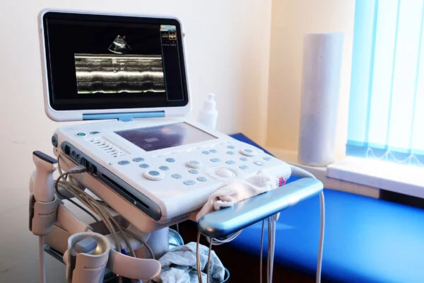
Detailed color Doppler ultrasound is an advanced imaging method used to evaluate vessels and blood flow. This technique uses ultrasound waves to display the direction and speed of blood flow inside the vessel in color. It provides high accuracy in detecting stenosis, insufficiency, or thrombus in both the arterial and venous systems.
In the diagnosis of venous insufficiency, deep vein thrombosis, or varicose veins, Doppler ultrasound forms the basis of diagnosis and treatment planning. During the examination, the probe is placed on the skin and sound waves reflect from the tissues to produce real-time images. The procedure is painless, radiation-free, and a safe diagnostic method.
Color Doppler ultrasound is particularly important for mapping the vessels of the lower extremities. Vessel diameter, valve function, and the direction of reflux are assessed in detail. This enables accurate planning before endovenous treatments or surgical interventions.
It is also used in the diagnosis of arterial stenosis, aneurysm, or organ perfusion disorders. Regular Doppler follow-ups help monitor vessel patency and circulatory efficiency after treatment. Having it performed by an experienced cardiovascular surgeon or radiologist increases the reliability of the result.
https://youtu.be/cZP6u8pU5Ugbr%20/

The operating principle of this technology is based on the “Doppler Effect.” Put simply, it’s the same effect where the siren of an approaching ambulance sounds higher-pitched and becomes lower-pitched as it moves away.
The logic is the same in medical imaging. The ultrasound device’s transducer, called a “probe,” sends sound waves that are inaudible to the human ear into your body. These sound waves strike the blood cells moving inside your vessels and are reflected back.
The device analyzes the change in frequency (pitch) of these returning sound waves. If blood cells are moving toward the probe, the frequency increases; if they are moving away, it decreases. By analyzing these frequency shifts within seconds, the device can measure the direction and speed of blood flow. To understand the severity of a vascular narrowing, the “speed” of blood is key for us.
During a Doppler examination, you see several information layers on the screen at the same time. Each is a different piece of the puzzle.
The main layers are:
The gray-scale image is the “anatomic map” of your tissues, organs, and vessels. Blood itself usually appears “black” on this image.
The color map is overlaid on this gray background. These colors indicate that there is blood flow in that region. The colors tell us the direction of flow: Red generally indicates blood moving toward the probe, and blue indicates blood moving away from the probe.
Here we should correct a very important misconception. The colors have nothing to do with “dirty” or “clean” blood (artery vs. vein). This is purely a coding based on the angle at which the probe is held. Sometimes these colors take on a “mosaic” appearance (green or yellow tones); this indicates turbulent flow in that area, which usually points to the segment just beyond a stenosis.
Spectral analysis is the most “mathematical” part of the exam. The rhythmic “whirring” sound you hear during the procedure comes from here. When a suspicious point is identified on the color map (for example a region with acceleration), the cursor is placed directly over that point. The device then plots the blood flow velocities at that single point as a “velocity–time graph.” This graph allows us to calculate the degree of stenosis as a percentage.
The quality and accuracy of a Doppler examination depend entirely on the experience of the operator and how well the device settings are adjusted. From the outside it may look like the probe is simply being moved, but in the background dozens of parameters are constantly being tuned. Incorrect settings can make a healthy vessel look diseased or a severe stenosis appear normal.
Some of the most frequently adjusted parameters are:
Gain is like the device’s “volume” control. If we turn it up too much, you get a “color confetti” effect—colors appearing even where there is actually no flow—resulting in a noisy image. If we turn it down too much, we cannot see slow flow (for example, weak flow at the edge of a thrombus) and the vessel may appear occluded.
Scale is like the device’s “speed dial.” To examine fast-flowing arteries, we need a “high gear,” and for slow-flowing veins, we need a “low gear.” If we look at an artery in a low gear, we get an image called “aliasing,” where colors wrap and mix. This is not an error; in fact it is a clue for us. This wrapping indicates that blood in that region is flowing much faster than expected—often a sign of significant stenosis.
The angle is critical for accurate velocity measurement. To measure blood flow velocity correctly, the sound beam must strike the vessel at a certain angle (ideally below 60 degrees). It’s like how a traffic officer must hold a radar gun parallel to the roadway to measure a car’s speed accurately.

Yes—and they have very important meanings. An experienced specialist can glean preliminary information about vascular health just by listening to the sound signature (waveform) on Spectral Doppler. Each vessel type has its own “sound signature”:
The main sound types are:
High-resistance sound is heard in healthy arm and leg arteries.
Low-resistance sound is characteristic of organs such as the brain and kidneys, which need continuous blood supply throughout the cardiac cycle (both systole and diastole). Pathological sounds are warning signs. For example, as blood passes through a stenosis it loses energy, so distal flow becomes dampened.
Peripheral arterial disease (PAD)—narrowing and calcification of leg arteries—is also known as “window-shopping disease.” A person starts walking, after a certain distance (e.g., 100 meters) cramps develop in the leg, they must stop, and the pain resolves with rest. This is a classic sign that the leg is not receiving enough blood.
Doppler ultrasound is the primary and most important tool both for diagnosing this disease and for planning treatment. Its purpose is to determine in which vessel, over what length, and to what severity a stenosis exists.
The exam starts with the patient lying on their back. Beginning at the groin, the “frog-leg position” may be requested (knee flexed and externally rotated). This position provides the ideal angle to visualize the groin and thigh vessels.
Step by step, the entire arterial system is surveyed. The main checkpoints are:
Calf arteries (if there is significant upstream stenosis or if there is a foot/leg ulcer)
In each segment, first the vessel wall is assessed on gray-scale imaging (is there calcification?), then a color Doppler flow map is obtained (is there a region where colors accelerate and become mosaic-like?). If a suspicious point is found, its velocity is measured with Spectral Doppler.
This is entirely based on velocity measurement. It’s a simple rule of physics. If you partially cover the end of a garden hose with your finger, the water jets out faster and with more force. The same applies to vessels: blood must accelerate when passing through a narrow segment.
A “ratio” is used to determine the severity. The velocity within the stenosis is compared to the velocity in the normal segment just proximal to it.
Classification is generally as follows:
Deep Vein Thrombosis (DVT) is the occlusion of the deep veins of the legs by a blood clot. If this clot breaks off and travels to the lungs (pulmonary embolism), it can be life-threatening. It is the most important cause of sudden leg swelling, pain, and redness. Doppler ultrasound is the gold standard method for diagnosing DVT.
This is the most fundamental and reliable method for diagnosing DVT. During the exam, the specialist gently presses the ultrasound probe over the vein.
Throughout the exam—from the groin to behind the knee and even into the calf—this “press–release” maneuver is repeated every 1–2 cm. Finding a segment that does not collapse is sufficient for diagnosing DVT. Additionally, with Color Doppler, we can see whether the thrombus completely occludes the vein or only partially. If a thrombus is present, color flow is either absent (complete occlusion) or bypasses around the thrombus (partial occlusion).
This is vital for treatment planning—and yes, Doppler can largely distinguish this. Their appearances and features differ.
Acute (New) Thrombus features:
Chronic (Old) Thrombus features:
This distinction is crucial for treatment. New thrombus responds best to anticoagulant therapy. Also, if a patient had DVT 6 months ago and the leg is swollen again today, the only way to determine whether this is a new (acute) clot or swelling due to damage left by the old (chronic) clot is via this assessment.
One of Doppler ultrasound’s greatest advantages is that it is “real-time.” So it is used not only for diagnosis but also as our “eyes” during treatment itself. This increases safety and success rates.
Main uses include:
For example, in intensive care when placing a catheter in the neck, rather than inserting a needle “blindly,” ultrasound allows us to visualize the needle tip, the target vein, and the adjacent carotid artery simultaneously. With Color Doppler we can target the vein (often coded blue) and actively avoid the artery (often coded red). This makes the procedure much safer and prevents unwanted injuries and complications.
Each imaging method has its place and they complement each other. The choice depends on the patient’s condition and what we are looking for.
Doppler Ultrasound (DUS)
Computed Tomography Angiography (CTA)
Magnetic Resonance Angiography (MRA)
Detailed Color Doppler Ultrasound is used to evaluate vascular occlusions, varicose veins, thrombi, or organ perfusion disorders. In pregnancy, it is preferred to assess blood flow to the baby and placenta.
In pregnancy, evaluation of the deep venous system can be safely performed in all trimesters. Additionally, at 6–8 weeks postpartum, a detailed color Doppler examination can be used to plan treatment.
During the procedure, a special gel is applied and the ultrasound probe is placed on the vessel or organ. The device visualizes blood flow and vessel structure in color using sound waves. The procedure is painless and contains no radiation.
Vascular occlusions, thrombi, varicose veins, venous insufficiency, reflux, and leak problems can be identified.
Unlike conventional ultrasound, it shows blood flow inside the vessels in color and motion. Thus, vessel structure, direction, and flow velocity are analyzed in detail, increasing diagnostic accuracy.
No, this method is completely safe. It uses sound waves, so there is no radiation. No adverse effects have been reported in pregnant women or babies.
Depending on the region examined, it takes on average 20–40 minutes. By mapping the vessels in both legs, problematic veins are identified and a treatment plan is made.
For some intra-abdominal vessel studies, fasting may be required. However, in most Doppler ultrasound applications, no special preparation is necessary. Wearing comfortable clothing helps the procedure.
Results can usually be obtained immediately after the examination. The images are evaluated and a report is prepared. A phlebologist can dictate the report during the scan and present it to you right away. However, especially in hospitals, reporting may be done later.
Price varies depending on the number of regions examined, the technology of the device used, the location of the center, and the experience of the specialist physician.

Varicose vein treatment prices are determined directly according to a treatment plan tailored specifically for [...]
Read More ➜
A successful varicose vein treatment depends on two critical phases. The most important step before [...]
Read More ➜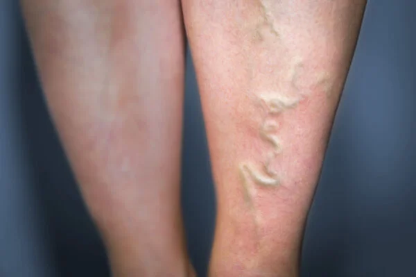
After varicose vein treatment, there is a risk of recurrence, but this risk can be [...]
Read More ➜